Translated from Greek it sounds like “white blood cells”. They are also called white blood cells. They capture and neutralize bacteria, so the main role of white blood cells is to protect the body from disease.
Antonina Kamyshenkova / “Health-Info”
When your white blood cell count changes
Slight fluctuations in your white blood cell count are completely normal. But the blood reacts very sensitively to any negative processes in the body, and in a number of diseases the level of white blood cells changes dramatically. A low level (below 4000 per 1 ml) is called leukopenia, and it can be a consequence, for example, of poisoning with various poisons, radiation, a number of diseases (typhoid fever), and also develop in parallel with iron deficiency anemia. And an increase in white blood cells - leukocytosis - can also be a consequence of certain diseases, for example, dysentery.
If the number of white blood cells increases sharply (up to hundreds of thousands in 1 ml), then this means leukemia - acute leukemia. With this disease, the process of hematopoiesis in the body is disrupted, and many immature white blood cells are formed - blasts, which cannot fight microorganisms. This is a deadly disease, and if left untreated, the patient is at risk.
Hello! I decided to write an article about white blood cells because many students confuse them. Ignorance of the types of leukocytes can bring a lot of problems in fundamental and clinical subjects. This is a very important material, without which it is impossible to understand even banal inflammation.
Let's immediately define a couple of basic things:
- Leukocytes are the formed elements of blood. Blood consists of formed elements and plasma. Formed elements include red blood cells, platelets and leukocytes. It is precisely the latter that we will deal with today;
- Leukocytes are white blood cells, at least that’s what people call them. We will call them formed elements, and not corpuscles, it is more correct;
- Leukocytes are our immunity. More precisely, our immunity is the blood-forming organs and the blood itself. But the direct immune response to any intervention in our body will be carried out precisely by leukocytes. That's why they are so interesting.
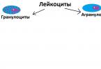
Two types of leukocytes
All leukocytes are divided into granular (they are also called granulocytes) and smooth (they are also called agranulocytes).
Granular leukocytes carry a bunch of “grains” within themselves. Each grain contains many aggressive biologically active substances. Imagine a soldier with a bunch of bombs, each filled with sulfuric acid - this is what granulocytes look like. Granulocyte “bombs” do not contain sulfuric acid, but there are many other aggressive substances and enzymes that literally corrode organic matter.
Smooth leukocytes are a completely different matter. Scientifically, they are called “agranulocytes,” that is, “cells without granules.” They do not carry any projectiles, however, they are also excellent at fighting, they just act more sophisticated and cunning. We will also talk about this.
For now, let’s draw the main division of leukocytes:

Granulocytes
Great, now let's look at granular leukocytes (ie granulocytes). They are divided into three main subclasses: neutrophils, basophils and eosinophils.
These complex names come from the coloring. If we stain using the Romanovsky-Giemsa method, granular leukocytes can be stained blue, that is, the main dye, and then we will call them basophils. Other white blood cells will stain only with the acidic eosinophil dye (which is red), and we will call them eosinophils. Still others are slightly colored by both, that is, they are neutral - neither basic nor acidic. Therefore we will call them neutrophils.
Neutrophils, despite their name, are not at all a neutral party in the event of an attack on our body by foreign agents. They frantically rush at the enemy, for example, at bacteria, tear them apart and devour them. True, they themselves die. Such berserkers are part of our blood. By the way, the pus that you can observe with severe inflammation, for example, of the tonsils or soft tissues around the fingernail, is nothing more than debris from dead neutrophils.

An important feature of neutrophils is that they non-specific in relation to foreign agents. In general, neutrophils are one of the main components of nonspecific immunity. It doesn’t matter to them what is in front of them - a bacterium, a protozoan, a foreign tissue or a splinter - they will try to tear and absorb everything.

An important thing is that eosinophils and especially neutrophils are pronounced phagocytes (“phagos” - devour), that is, cells that devour other cells. They are usually called microphages, as they are quite small.
Basophils. The last type of granular leukocyte we are considering. Basophil granules contain substances that help allergic reactions. This is, of course, histamine, as well as serotonin and leukotrienes. Basophils are found in extremely small quantities in a healthy body. They become more numerous in cases of violent immediate allergic reactions, as well as in lymphomas, leukemia and some autoimmune diseases.
Let's reflect on our diagram what we learned:

Agranulocytes
Let's move on to the second half of the classification. Now we will consider agragulocytes, that is, smooth leukocytes devoid of granular granules. First of all, we divide agranulocytes into two groups: monocytes and lymphocytes.

Monocytes
Monocytes are very large cells. Their main task is phagocytosis, that is, devouring, like neutrophils.
However, monocytes are quite large eater cells, so we will call them macrophages. Moreover, in addition to phagocytosis, monocytes are able to synthesize and secrete a whole class of substances called monokines. Monokines include interleukins 1, 6 (stimulate the production of antibodies, more on them later), tumor necrosis factor and others.
Another important feature is that monocytes are able to inhabit a variety of tissues and ripen there to specialized cells called tissue macrophages. This is a very mature cell that “devours” enemies only within a certain tissue. If a monocyte settles in the liver tissue and matures there, it is called a Kupffer cell. If it decides to settle in bone tissue, it becomes an osteoclast. In the lungs this will be an alveolar macrophage.
Finally, monocytes and macrophages are capable of not only engulfing bacteria or cells infected with a virus - they can also expose its fragments onto its surface so that it can be recognized by T-helpers, which we’ll talk about later.
We sign the final stage of monocyte development and move on.

Lymphocytes
So, we already know that smooth leukocytes (agranulocytes) are divided into monocytes and lymphocytes. We have already discussed monocytes, now let's talk about lymphocytes.
Lymphocytes are the most important cells of the immune system. They are the ones who carry out full specific immune response, protecting us from numerous threats both from outside (viruses, bacteria) and from inside us (cells). Before we analyze them in detail, it is necessary to immediately divide them into two large groups - T-lymphocytes and B-lymphocytes. There is also a conditionally independent group, which is extremely interesting. But first things first.

T lymphocytes
The first stages of maturation in these lymphocytes (like all blood cells in general) take place in the red bone marrow. And their ripening to full-fledged cells in the thymus is completed (the thymus is the thymus gland). That is why they are called T-lymphocytes, that is, “Thymus-Lymphocytes”. T lymphocytes are the main representatives of cellular immunity.
That is, they start working when there is a danger to the body - cell(bacteria, cancer cell), or something that has gotten inside a body cell (for example, a virus).
So, T lymphocytes are the main representatives of the immune cell line. They are divided into at least 4 groups:
- Killer T cells. The real killers. They are the ones who kill bacteria and cells infected by the virus. Killer T cells also attack foreign tissue transplanted from another person. T-killers can also attack cells with signs of tumor degeneration, but the main characters of this story are waiting for us ahead;
- T helper cells, from the English “help” - “help”. Extremely important lymphocytes. T-helpers coordinate the coordinated work of cellular and humoral immunity. They react to the situation inside the body and quickly give orders to certain cells to divide, attack or mature. By the way, HIV infection primarily affects these cells, as well as tissue macrophages.
- T-suppressors, from the English “suppress” - to suppress. These T lymphocytes suppress the activity of killer T cells and helper T cells. If the T-suppressors do not work enough, the killer and helper cells will become so divergent that they will begin to attack their own tissues. T-suppressors make sure that this does not happen.
- T-memory. This long-lived lymphocyte retains information about what foreign agents the body has encountered previously. Thanks to T-memory, the immune response during a second encounter, for example, with a virus, can be carried out much faster than the first time.
Let's draw T-lymphocytes in our diagram, all four types, and move on.

B lymphocytes
Remember when we said that T-lymphocytes mature in the thymus, which is why they are called that? So, B-lymphocytes mature in the red bone marrow and that's why they are called...B-lymphocytes? In fact, they were first discovered in birds, in an organ called the bursa of Fabricius. "Bag" in English is "bursa", which is where the "B" comes from.
The main function of all B-lymphocytes is the production of antibodies, that is immunoglobulins. Immunoglobulin is a special protein that binds to a foreign agent and neutralizes it. Often immunoglobulins bind to a variety of poisons and bacterial breakdown products in order to neutralize them.
Immunoglobulins are one of the most important components of humoral immunity. Remember when we said that cellular immunity works when the foreign agent is a cell? So, humoral immunity works when a foreign agent is substance, soluble in liquid, or liquid. You can easily remember this rule, because in Greek “humor” means “liquid”.
An important point is that in order to begin producing immunoglobulin, the B lymphocyte must transform into a plasma cell. It is plasma cells that produce antibodies (immunoglobulins). In order for a B lymphocyte to turn into a plasma cell, several signals are required, for example, from the T helper cells that we talked about.
What's even more interesting is that the plasma cell is capable of converting back into a B lymphocyte once its mission of producing immunoglobulin is completed.
Not exactly the topic of this article, but it is important: cellular and humoral immunity are separated in our classifications for better understanding, but in reality they tend to work together under the guidance of T helper cells and other immune cells.
We add the B-line to our diagram and proceed to the last part of the story about leukocytes.

NK lymphocytes
Remember, we said that we have an extremely interesting independent group of lymphocytes that do not belong to either the T or B lineages? So, we are talking about the so-called NK lymphocytes, also called “natural killers”. "NK" - from the abbreviation "Natural Killers", that is, " natural killers«.

There are very unusual lymphocyte cells in our blood. Their receptor structure does not allow them to be classified as T- or B-lines. They are able to recognize various cells of the body, identifying those that are affected by a virus or are on the way to degenerate into a cancer cell.
Once again - NK lymphocytes recognize And kill cancer cells and virus-infected cells. Moreover, they can recognize cancer cells at a more subtle level than T-lymphocytes. This is a very important layer of anti-cancer defense, in fact.
When an NK lymphocyte notices a suspiciously altered cell in the body, it launches a massive attack. NK literally perforates a potentially cancerous cell in several places at once, after which water and sodium begin to flow into it in huge quantities. Next, the potentially cancerous cell dies from membrane rupture.
It has always seemed to me a great injustice that on histological plates, various hematopoietic schemes, even in the hematological sections of textbooks, NK cells are ignored. That is, they are not mentioned at all, or they simply write that they exist. In my opinion, this is wrong - for a moment, we are talking about immune cells that protect us from cancer. But in our table they will take their rightful place:

I hope I didn't confuse you too much. If you suddenly do not understand some stage of my scheme, write about it in the comments, I will be happy to tell you everything. I plan to make a couple more posts about hematopoiesis in the red bone marrow and about leukemia. However, without mastering this material about leukocytes, you will not understand anything, so try to thoroughly understand these diagrams. It is best, of course, to draw the same ones and sign special signs next to each class.
I would like to dedicate this manual to the memory of Elena Borisovna Rodzaevskaya, a brilliant teacher and an amazing person. She invested a lot in me and thanks to her I have some knowledge that I share with you, dear readers. Elena Borisovna believed in me when practically no one did. I am very proud that I had the opportunity to be her student.
Blood circulates continuously in the system of blood vessels. It performs very important functions in the body: respiratory, transport, protective and regulatory, ensuring the constancy of the internal environment of our body.
Blood is one of the connective tissues, which consists of a liquid intercellular substance that has a complex composition. It includes plasma and cells suspended in it, or the so-called formed elements of blood: leukocytes, erythrocytes and platelets. It is known that in 1 mm 3 of blood there are from 5 to 8 thousand leukocytes, 4.5 to 5 million erythrocytes, and 200 to 400 thousand platelets.
The amount of blood in the body of a healthy person is approximately 4.5 to 5 liters. Plasma occupies 55-60% of the volume, and 40-45% of the total volume remains for formed elements. Plasma is a translucent yellowish liquid that contains water (90%), organic and mineral substances, vitamins, amino acids, hormones, and metabolic products.
Structure of leukocytes
Red blood cells

There are red blood cells and white blood cells in the blood. Their structure and functions are different from each other. An erythrocyte is a cell that has the shape of a biconcave disc. It does not contain a nucleus, and most of the cytoplasm is occupied by a protein called hemoglobin. It consists of an iron atom and a protein part and has a complex structure. Hemoglobin carries oxygen in the body.
Red blood cells appear in the bone marrow from erythroblast cells. Most red blood cells are biconcave in shape, but the rest may vary. For example, they can be spherical, oval, bitten, cup-shaped, etc. It is known that the shape of these cells can be disrupted due to various diseases. Each red blood cell stays in the blood for 90 to 120 days, and then dies. Hemolysis is the phenomenon of destruction of red blood cells, which occurs mainly in the spleen, as well as in the liver and blood vessels.
Platelets

The structure of leukocytes and platelets is also different. Platelets do not have a nucleus; they are small oval or round cells. If these cells are active, outgrowths form on them, they resemble a star. Platelets appear in the bone marrow from the megakaryoblast. They “work” for only 8 to 11 days, then they die in the liver, spleen or lungs.
Very important. They are able to maintain the integrity of the vascular wall and restore it in case of damage. Platelets form a clot and thereby stop bleeding.
Human blood is a liquid substance consisting of plasma and formed elements, or blood cells, suspended in it, which make up approximately 40-45% of the total volume. They are small in size and can only be seen under a microscope.
There are several types of blood cells that perform specific functions. Some of them function only within the circulatory system, others go beyond its boundaries. What they have in common is that they are all formed in the bone marrow from stem cells, the process of their formation is continuous, and their lifespan is limited.
All blood cells are divided into red and white. The first are erythrocytes, which make up the majority of all cells, the second are leukocytes.
Platelets are also considered to be blood cells. These small platelets of blood are not actually full-fledged cells. They are small fragments separated from large cells - megakaryocytes.
Red blood cells are called red blood cells. This is the most numerous group of cells. They carry oxygen from the respiratory organs to the tissues and take part in the transport of carbon dioxide from the tissues to the lungs.
The place of formation of red blood cells is the red bone marrow. They live for 120 days and are destroyed in the spleen and liver.
They are formed from precursor cells - erythroblasts, which, before becoming an erythrocyte, go through different stages of development and divide several times. Thus, up to 64 red blood cells are formed from the erythroblast.

Red blood cells lack a nucleus and are shaped like a disk concave on both sides, the diameter of which is on average about 7-7.5 microns, and the thickness at the edges is 2.5 microns. This shape increases the ductility required for passage through small vessels and the surface area for gas diffusion. Old red blood cells lose their plasticity, which is why they linger in the small vessels of the spleen and are destroyed there.
Most red blood cells (up to 80%) have a biconcave spherical shape. The remaining 20% may have another: oval, cup-shaped, simple spherical, crescent-shaped, etc. Violation of the shape is associated with various diseases (anemia, deficiency of vitamin B 12, folic acid, iron, etc.).

Most of the cytoplasm of the red blood cell is occupied by hemoglobin, consisting of protein and heme iron, which gives the blood its red color. The non-protein part consists of four heme molecules with an Fe atom in each. It is thanks to hemoglobin that the red blood cell is able to carry oxygen and remove carbon dioxide. In the lungs, an iron atom binds with an oxygen molecule, hemoglobin turns into oxyhemoglobin, which gives the blood a scarlet color. In tissues, hemoglobin gives up oxygen and adds carbon dioxide, turning into carbohemoglobin, as a result the blood becomes dark. In the lungs, carbon dioxide is separated from hemoglobin and removed by the lungs to the outside, and the incoming oxygen is again associated with iron.
In addition to hemoglobin, the cytoplasm of the erythrocyte contains various enzymes (phosphatase, cholinesterase, carbonic anhydrase, etc.).
The erythrocyte membrane has a fairly simple structure compared to the membranes of other cells. It is an elastic thin mesh, which ensures rapid gas exchange.
On the surface of red blood cells there are different types of antigens that determine the Rh factor and blood group. The Rh factor can be positive or negative depending on the presence or absence of the Rh antigen. The blood group depends on which antigens are on the membrane: 0, A, B (the first group is 00, the second is 0A, the third is 0B, the fourth is AB).
In the blood of a healthy person, there may be small amounts of immature red blood cells called reticulocytes. Their number increases with significant blood loss, when replacement of red cells is required and the bone marrow does not have time to produce them, so it releases immature ones, which are nevertheless capable of performing the functions of red blood cells in transporting oxygen.
Leukocytes are white blood cells whose main task is to protect the body from internal and external enemies.
They are usually divided into granulocytes and agranulocytes. The first group is granular cells: neutrophils, basophils, eosinophils. The second group does not have granules in the cytoplasm; it includes lymphocytes and monocytes.

This is the most numerous group of leukocytes - up to 70% of the total number of white cells. Neutrophils got their name due to the fact that their granules are stained with dyes with a neutral reaction. Its grain size is fine, the granules have a purple-brownish tint.

The main task of neutrophils is phagocytosis, which consists in capturing pathogenic microbes and tissue breakdown products and destroying them inside the cell with the help of lysosomal enzymes found in granules. These granulocytes fight mainly against bacteria and fungi and to a lesser extent against viruses. Pus consists of neutrophils and their remains. During the breakdown of neutrophils, lysosomal enzymes are released and soften nearby tissues, thus forming a purulent focus.
A neutrophil is a rounded nuclear cell, reaching a diameter of 10 microns. The core may have the form of a rod or consist of several segments (from three to five) connected by strands. An increase in the number of segments (up to 8-12 or more) indicates pathology. Thus, neutrophils can be band or segmented. The first are young cells, the second are mature. Cells with a segmented nucleus make up up to 65% of all leukocytes, and band cells in the blood of a healthy person make up no more than 5%.

The cytoplasm contains about 250 types of granules containing substances through which the neutrophil performs its functions. These are protein molecules that affect metabolic processes (enzymes), regulatory molecules that control the work of neutrophils, substances that destroy bacteria and other harmful agents.
These granulocytes are formed in the bone marrow from neutrophilic myeloblasts. A mature cell stays in the brain for 5 days, then enters the blood and lives here for up to 10 hours. From the vascular bed, neutrophils enter the tissues, where they remain for two to three days, then they enter the liver and spleen, where they are destroyed.
There are very few of these cells in the blood - no more than 1% of the total number of leukocytes. They have a round shape and a segmented or rod-shaped nucleus. Their diameter reaches 7-11 microns. Inside the cytoplasm there are dark purple granules of varying sizes. They got their name due to the fact that their granules are colored with dyes with an alkaline, or basic, reaction. Basophil granules contain enzymes and other substances involved in the development of inflammation.

Their main function is the release of histamine and heparin and participation in the formation of inflammatory and allergic reactions, including the immediate type (anaphylactic shock). In addition, they can reduce blood clotting.

They are formed in the bone marrow from basophilic myeloblasts. After maturation, they enter the blood, where they remain for about two days, then go into the tissues. What happens next is still unknown.
These granulocytes make up approximately 2-5% of the total white cells. Their granules are stained with an acidic dye, eosin.
They have a rounded shape and a slightly colored core, consisting of segments of the same size (usually two, less often three). Eosinophils reach 10-11 microns in diameter. Their cytoplasm is painted pale blue and is almost invisible among the large number of large round granules of yellow-red color.

These cells are formed in the bone marrow, their precursors are eosinophilic myeloblasts. Their granules contain enzymes, proteins and phospholipids. A mature eosinophil lives in the bone marrow for several days, after entering the blood it remains in it for up to 8 hours, then moves to tissues that have contact with the external environment (mucous membranes).

These are round cells with a large nucleus occupying most of the cytoplasm. Their diameter is 7 to 10 microns. The kernel is round, oval or bean-shaped and has a rough structure. Consists of lumps of oxychromatin and basiromatin, resembling blocks. The core can be dark purple or light purple, sometimes it contains light inclusions in the form of nucleoli. The cytoplasm is colored light blue; around the nucleus it is lighter. In some lymphocytes, the cytoplasm has azurophilic granularity, which turns red when stained.

Two types of mature lymphocytes circulate in the blood:
- Narrow plasma. They have a rough dark purple nucleus and a narrow blue rim of cytoplasm.
- Wide-plasma. In this case, the kernel has a paler color and bean-shaped shape. The rim of the cytoplasm is quite wide, gray-blue in color, with rare ausurophilic granules.
From atypical lymphocytes in the blood you can find:
- Small cells with barely visible cytoplasm and a pyknotic nucleus.
- Cells with vacuoles in the cytoplasm or nucleus.
- Cells with lobed, kidney-shaped, jagged nuclei.
- Bare kernels.
Lymphocytes are formed in the bone marrow from lymphoblasts and undergo several stages of division during the process of maturation. Its complete maturation occurs in the thymus, lymph nodes and spleen. Lymphocytes are immune cells that mediate immune responses. There are T-lymphocytes (80% of the total) and B-lymphocytes (20%). The former matured in the thymus, the latter in the spleen and lymph nodes. B lymphocytes are larger in size than T lymphocytes. The lifespan of these leukocytes is up to 90 days. Blood for them is a transport medium through which they enter tissues where their help is required.
The actions of T-lymphocytes and B-lymphocytes are different, although both take part in the formation of immune reactions.
The former are engaged in the destruction of harmful agents, usually viruses, through phagocytosis. The immune reactions in which they participate are nonspecific resistance, since the actions of T lymphocytes are the same for all harmful agents.
According to the actions they perform, T-lymphocytes are divided into three types:
- T-helpers. Their main task is to help B-lymphocytes, but in some cases they can act as killers.
- T-killers. Destroy harmful agents: foreign, cancerous and mutated cells, infectious agents.
- T-suppressors. Inhibit or block overly active reactions of B-lymphocytes.
B-lymphocytes act differently: against pathogens they produce antibodies - immunoglobulins. This happens as follows: in response to the actions of harmful agents, they interact with monocytes and T-lymphocytes and turn into plasma cells that produce antibodies that recognize the corresponding antigens and bind them. For each type of microbe, these proteins are specific and are capable of destroying only a certain type, therefore the resistance that these lymphocytes form is specific, and it is directed primarily against bacteria.

These cells provide the body's resistance to certain harmful microorganisms, which is commonly called immunity. That is, having encountered a harmful agent, B-lymphocytes create memory cells that form this resistance. The same thing - the formation of memory cells - is achieved by vaccinations against infectious diseases. In this case, a weak microbe is introduced so that the person can easily survive the disease, and as a result, memory cells are formed. They can remain for life or for a certain period, after which the vaccination must be repeated.
Monocytes are the largest of the leukocytes. Their number ranges from 2 to 9% of all white blood cells. Their diameter reaches 20 microns. The monocyte nucleus is large, occupies almost the entire cytoplasm, can be round, bean-shaped, mushroom-shaped, or butterfly-shaped. When stained it turns red-violet. The cytoplasm is smoky, bluish-smoky, less often blue. It usually has an azurophilic fine grain size. It may contain vacuoles (voids), pigment grains, and phagocytosed cells.

Monocytes are produced in the bone marrow from monoblasts. After maturation, they immediately appear in the blood and remain there for up to 4 days. Some of these leukocytes die, some move into the tissue, where they mature and turn into macrophages. These are the largest cells with a large round or oval nucleus, blue cytoplasm and a large number of vacuoles, which is why they appear foamy. The lifespan of macrophages is several months. They can be constantly in one place (resident cells) or move around (wandering cells).

Monocytes form regulatory molecules and enzymes. They are able to form an inflammatory response, but can also inhibit it. In addition, they participate in the wound healing process, helping to speed it up, and promote the restoration of nerve fibers and bone tissue. Their main function is phagocytosis. Monocytes destroy harmful bacteria and inhibit the proliferation of viruses. They are able to carry out commands, but cannot distinguish between specific antigens.
These blood cells are small, anucleate plates and can be round or oval in shape. During activation, when they are near the damaged vessel wall, they form outgrowths, so they look like stars. Platelets contain microtubules, mitochondria, ribosomes, and specific granules containing substances necessary for blood clotting. These cells are equipped with a three-layer membrane.

Platelets are produced in the bone marrow, but in a completely different way than other cells. Blood plates are formed from the largest cells of the brain - megakaryocytes, which, in turn, were formed from megakaryoblasts. Megakaryocytes have a very large cytoplasm. After the cell matures, membranes appear in it, dividing it into fragments that begin to separate, and thus platelets appear. They leave the bone marrow into the blood, stay in it for 8-10 days, then die in the spleen, lungs, and liver.

Blood plates can have different sizes:
- the smallest are microforms, their diameter does not exceed 1.5 microns;
- normoforms reach 2-4 microns;
- macroforms – 5 microns;
- megaloforms – 6-10 microns.
Platelets perform a very important function - they participate in the formation of a blood clot, which closes the damage in the vessel, thereby preventing blood from leaking out. In addition, they maintain the integrity of the vessel wall and promote its rapid recovery after damage. When bleeding begins, platelets adhere to the edge of the injury until the hole is completely closed. The adhered plates begin to break down and release enzymes that affect the blood plasma. As a result, insoluble fibrin threads are formed, tightly covering the injury site.
Conclusion
Blood cells have a complex structure, and each type performs a specific job: from transporting gases and substances to producing antibodies against foreign microorganisms. Their properties and functions have not been fully studied to date. For normal human life, a certain amount of each type of cell is necessary. Based on their quantitative and qualitative changes, doctors have the opportunity to suspect the development of pathologies. The composition of the blood is the first thing that a doctor studies when treating a patient.
The main sphere of action of leukocytes is protection. They play a major role in the specific and nonspecific protection of the body from external and internal pathogenic agents, as well as in the implementation of typical pathological processes.
All types of leukocytes are capable of active movement and can pass through the capillary wall and penetrate into the intercellular space, where they absorb and digest foreign particles. This process is called phagocytosis, and the cells that carry it out are phagocytes.
If a lot of foreign bodies have entered the body, then the phagocytes, absorbing them, greatly increase in size and are eventually destroyed. This releases substances that cause a local inflammatory reaction, which is accompanied by swelling, fever and redness of the affected area.
Substances that cause an inflammatory reaction attract new leukocytes to the site of foreign body penetration. By destroying foreign bodies and damaged cells, leukocytes die in large quantities. Pus, which forms in tissues during inflammation, is an accumulation of dead leukocytes.
The blood of an adult contains 1000 times less leukocytes than red blood cells, and on average their number is 4-9·10 9 /l. In newborn children, especially in the first days of life, the number of leukocytes can vary greatly from 9 to 30·10 9 /l. In children aged 1-3 years, the number of leukocytes in the blood fluctuates between 6.0-17.0·10 9 /l, and at 6-10 years old within 6.0-11.0·10 9 /l.
An increase in the total absolute number of leukocytes per unit volume above the upper limit of normal is called absolute leukocytosis, and decreasing it below the lower limit - absolute leukopenia.
Leukocytosis
True leukocytosis occurs when the formation of leukocytes increases and their release from the bone marrow. If the increase in the content of leukocytes in the blood is associated with the entry into circulation of those cells that are under normal conditions attached to the inner surface of the vessels, such leukocytosis is called redistributive.
It is the redistribution of leukocytes that explains the fluctuations during the day. Thus, the number of leukocytes usually increases slightly in the evening, as well as after eating.
Physiological leukocytosis observed in the premenstrual period, in the second half of pregnancy, 1-2 weeks after delivery.
Physiological redistributive leukocytosis can be observed after eating, after physical or emotional stress, exposure to cold or heat.
Leukocytosis as a pathological reaction most often indicates an infectious or aseptic inflammatory process in the body. In addition, leukocytosis is often detected in cases of poisoning with nitrobenzene, aniline, in the initial phase of radiation sickness, as a side effect of certain medications, as well as in malignant neoplasms, acute blood loss and many other pathological processes. In its most severe form, leukocytosis occurs in leukemia.
Leukopenia
Leukopenia can also be physiological (constitutional leukopenia) and pathological, redistributive and true.
Some causes of leukopenia:
Leukocytes are a collective concept introduced in the 19th century and retained for the sake of simplicity in contrasting “white blood - red blood.” According to modern data, leukocytes differ in origin, function and appearance. Some leukocytes are able to capture and digest foreign microorganisms (phagocytosis), while others can produce antibodies. As a result, there are several types of leukocyte division, the simplest of which is based on the presence/absence of specific granules in their cytoplasm.
Based on morphological characteristics, leukocytes stained according to Romanovsky-Giemsa have been traditionally divided into two groups since the time of Ehrlich:
- granular leukocytes, or granulocytes- cells that have large segmented nuclei and exhibit a specific granularity of the cytoplasm; depending on the ability to perceive dyes, they are divided into neutrophilic, eosinophilic and basophilic;
- non-granular leukocytes, or agranulocytes- cells that do not have a specific granularity and contain a simple non-segmented nucleus, these include lymphocytes and monocytes.
The ratio of different types of white cells, expressed as a percentage, is called the leukocyte formula.
The study of the number and ratio of leukocytes is an important step in the diagnosis of diseases.
Important contributions to the study of the protective properties of leukocytes were made by Ilya Mechnikov and Paul Ehrlich. Mechnikov discovered and studied the phenomenon of phagocytosis, and subsequently developed the phagocytic theory of immunity. Ehrlich is responsible for the discovery of various types of leukocytes. In 1908, the scientists were jointly awarded the Nobel Prize for their services.
- G. I. Nazarenko, A. A. Kishkun, “Clinical evaluation of laboratory research results,” Moscow, 2005.
- A. A. Kishkun “Guide to laboratory research methods” 2007
- Knipovich N. M. White blood cells // Encyclopedic Dictionary of Brockhaus and Efron: in 86 volumes (82 volumes and 4 additional). - St. Petersburg. , 1890-1907.
- White blood cells // Small encyclopedic dictionary of Brockhaus and Efron: in 4 volumes - St. Petersburg. , 1907-1909.
- Norm of leukocytes in the blood in children and adults
- Leukocytes. Encyclopedic Dictionary.
You can edit this article to add links to authoritative sources.
Place of formation of leukocytes
The number of leukocytes is an important indicator for diagnosing pathological conditions. The body constantly produces white blood cells, and their levels in the blood can change throughout the day. How are these cells produced and what role do they play in the human body?
Place of formation of leukocytes
What are leukocytes
Several types of formed elements float in the blood, which support the health of the whole organism. White cells that have a nucleus inside are called leukocytes. Their peculiarity is the ability to penetrate the capillary wall and enter the intercellular space. It is there that they find foreign particles and absorb them, normalizing the vital activity of the cells of the human body.
Leukocytes include several types of cells that differ slightly in origin and appearance. The most popular division is based on morphological characteristics.
The ratio of these cells is the same in all healthy people and is expressed by the leukocyte formula. By changing the number of any type of cells, doctors draw conclusions about the nature of the pathological process.
What are leukocytes
Important: it is leukocytes that maintain human health at the proper level. Most infections that enter the human body are asymptomatic due to a timely immune response.
Functions of leukocytes
The importance of leukocytes is explained by their participation in the immune response and protecting the body from any foreign agents. The main functions of white cells are as follows:
- Antibody production.
- Absorption of foreign particles – phagocytosis.
- Destruction and removal of toxins.
Each type of leukocyte is responsible for certain processes that help in carrying out the main functions:
- Eosinophils. They are considered the main agents for the destruction of allergens. They participate in the neutralization of many foreign components that have a protein structure.
- Basophils. They accelerate the healing process at the site of inflammation, due to the presence of heparin in its structure. Updated every 12 hours.
- Neutrophils. Participate directly in phagocytosis. Capable of penetrating into the intercellular fluid and into the cell where the microbe lives. One such immune cell can digest up to 20 bacteria. Fighting microbes, the neutrophil dies. Acute inflammation provokes a sharp production of such cells by the body, which is immediately reflected in the leukocyte formula as an increased number.
- Monocytes. Helps neutrophils. They are more active if an acidic environment develops at the site of inflammation.
- Lymphocytes. They distinguish their own cells from foreign cells by their structure and participate in the production of antibodies. They live for several years. They are the most important component of immune defense.
Important: many doctors require you to do a clinical blood test before prescribing treatment. Viral and bacterial diseases cause different changes in the analysis, which makes it possible to make the correct diagnosis and prescribe the necessary medications.
Place of formation of leukocytes
All types of white blood cells are produced in the bone marrow, which is found inside the bones. It contains a huge number of immature cells, similar to those found in an embryo. From them, as a result of a complex multi-stage process, various hematopoietic cells are formed, including all types of leukocytes.
The transformation occurs as a result of the division of immature cells. With each stage they become more differentiated and designed to perform more specific functions. All stages, and there can be up to 9 of them, occur in the bone marrow. The exception is lymphocytes. To fully “grow up,” they will need to mature in the lymphoid organs.
Places of formation of leukocytes
Leukocytes accumulate in the bone marrow, and during the inflammatory process they enter the blood and reach the pathological focus. After fulfilling their purpose, the cells die, and the bone marrow forms new ones. Normally, only a small part of the body’s total leukocyte reserves floats in the bloodstream (up to 2%).
During the inflammatory process, all cells rush to the site of its localization. Neutrophil reserves for such emergency surges are located on the walls of blood vessels. It is this depot that allows the body to quickly respond to inflammation.
Lymphocytes can mature into T or B cells. The former regulate the production of antibodies, and the latter recognize foreign agents and neutralize them. Intermediate T cell development occurs in the thymus. The final maturation of lymphocytes occurs in the spleen and lymph nodes. It is there that they actively divide and turn into full-fledged immune defense. During inflammation, lymphocytes move to the nearest lymph node.
Important: the mechanism of leukocyte formation is very complex. Don't forget the importance of the spleen and other organs. For example, drinking alcohol has a negative effect on them.
Video - Leukocytes
Lack of white blood cells
Leukopenia in an adult is a condition when the number of leukocytes is below 4 * 10 9 / l. This may be caused by malignant diseases, exposure to radiation, vitamin deficiencies or problems with hematopoietic function.
Leukopenia leads to the rapid development of various infections and a decrease in the body's resistance. A person feels chills, body temperature rises, loss of strength and exhaustion appear. The body tries to compensate for the lack of defense cells, resulting in an enlarged spleen. This condition is very dangerous and requires identification of the cause and treatment.
What is leukopenia
Important: chronic fatigue or other conditions that have been bothering you for a long time should not be ignored. They often occur due to a decrease in the body's defenses.
Excess white blood cells
The number of leukocytes above 9*10 9 /l is considered to be above normal and is called leukocytosis. Physiological enlargement, which does not require treatment, can be caused by food intake, physical activity, certain hormonal surges (pregnancy, premenstrual period).
The following causes of leukocytosis lead to pathological conditions:
- Infectious diseases.
- Inflammatory processes of microbial and non-microbial etiology.
- Blood loss.
- Burns.
What is leukocytosis
Treatment for this condition may include the following groups of drugs:
- Antibiotics. They help eliminate the infection that caused leukocytosis and prevent complications.
- Steroid hormones. Quickly and effectively relieve inflammation, which leads to a decrease in the production of leukocytes.
- Antihistamines. Also help reduce inflammation.
The treatment tactics for any changes in the leukocyte formula depend on the cause that caused them.
Important: minor changes in the leukocyte formula may be temporary and even considered normal. Strong discrepancies with acceptable values or lack of changes during repeated analyzes should be alarming.
Children are taught about the importance of leukocytes at school. This topic is not an exaggeration. Good immunity ensures health and a good quality of life for every person. To determine the state of the immune system, you can take a blood test during the absence of illness. A competent doctor will help you interpret the results correctly.
Video - What does an increase in leukocytes in a blood test mean?
Leukocytes in the blood: determination of quantity, types and functions
During a general blood test, the absolute white blood cell count (WBC) is determined and the leukocyte formula is calculated. The latter determines the percentage and absolute content of lymphocytes, neutrophils, eosinophils, basophils and the percentage of monocytes. The percentage content in the decoding is designated as “%”, absolute content as “#”.
Leukocytes are heterogeneous in their morphology and priority protective function.
Standard indicators for determining the number of blood leukocytes do not have age (in persons over 18 years of age) and gender differences - 4.0 - 8.8x10⁻⁹/l.
The main reasons for a decrease in the level of blood leukocytes
The number of leukocytes in the blood changes under the influence of a number of external factors - seasonal, climatic, meteorological, as well as under various physiological conditions. Changes in the concentration of leukocytes can be recorded immediately after a meal, after physical activity, during pregnancy, and at certain phases of the menstrual cycle in women. Also, changes in the level of leukocytes in the blood can be affected by age-related changes and various pathologies.
A decrease in white blood cells is called leukopenia.
The main reasons for the decrease in white blood cells (leukopenia) are pathological and physiological changes:
Pathological factors reducing the level of leukocytes in the blood:
- insufficiency of hematopoiesis in the bone marrow due to the death of stem cells (the precursors of all blood cells) due to: exposure to ionizing radiation (radiation sickness); malignant neoplasms of blood cells (acute leukemia) or metastases in the case of a different tumor location; fatty degeneration of the bone marrow (aplastic anemia); exposure to certain medications (antitumor drugs, some antibiotics - for example, chloramphenicol, sulfonamides, pyrazolone derivatives, non-steroidal anti-inflammatory drugs, antiepileptic drugs, thyreostatics, etc.); exposure to certain chemicals (for example, benzene);
- some infections (influenza, brucellosis, measles, rubella, chickenpox, yellow fever, infectious mononucleosis);
- autoimmune diseases (systemic lupus erythematosus, etc.);
- anaphylactic shock;
- sepsis;
- diseases accompanied by hypersplenia (an increase in the size of the spleen and a decrease in the number of formed blood elements): liver damage due to cirrhosis, viral hepatitis; damage to the spleen in some congenital hemolytic anemias (thalassemia), some infections (malaria, typhoid fever, paratyphoid fever, tuberculosis); lymphogranulomatosis; amyloidosis, etc.
Physiological reasons for a decrease in leukocytes in the blood:
- starvation;
- hypotonic conditions;
- decrease in the overall tone of the body.
The main reasons for increased levels of leukocytes in the blood
An increase in the level of white blood cells in the blood is called leukocytosis.
The main reasons for the increase in white blood cells (leukocytosis) are pathological and physiological factors:
Pathological causes of increased leukocytes in the blood:
Physiological reasons for increased levels of leukocytes in the blood:
- after physical activity (myogenic leukocytosis);
- 2-3 hours after eating a meal, especially protein (this increase in leukocytes in the blood is called digestive leukocytosis);
- pregnancy, especially the second half;
- emotional stress;
- taking certain medications (steroids, adrenaline).
In the next section of the article, you will learn what types of leukocytes there are and what functions they perform.
What are the types of leukocytes in human blood, and what main functions do they perform?
There are the following types of blood leukocytes:
Granulocytes (cells containing specific granularity, the chemical composition of which determines the basic function of certain leukocytes).
The lifespan of this type of leukocyte is 9-13 days, usually for the first 5-6 days they are in the bone marrow, then they enter the bloodstream, where they circulate from several hours to two days, and then migrate to the tissues, where they carry out their functions.
Neutrophils - the main function of these leukocytes is to phagocytose (capture, neutralize and digest) most infectious agents. In addition, neutrophil granules contain substances that carry vitamin B12 and substances involved in the processes of reproduction, growth and regeneration of tissues of the human body.
Most of the neutrophils are found in the bone marrow (at varying degrees of maturation) and in body tissues, and in the peripheral blood there are only a small number of band (which belong to the class of maturing cells) and segmented - mature neutrophils. The lifespan of neutrophils is short - only a few days.
Eosinophilic leukocytes in their granules contain substances that neutralize biologically active substances released in the human body when allergic reactions of the immediate type occur (for example, with anaphylactic shock or Quincke's edema, requiring emergency medical measures) and delayed type (food and drug dermatitis).
Eosinophils are present in very small quantities in the blood; they usually accumulate in tissues in contact with the external environment (skin, lungs, gastrointestinal and urogenital tracts). Moreover, for this group of cells there is a clear daily rhythm of fluctuations in their number in the blood: at night, the maximum number of eosinophils in the blood is observed, and the minimum during the day. The lifespan of eosinophils is up to 8-12 days.
Basophils perform several functions: in addition to phagocytosis, which is characteristic of all granulocytes, they also maintain blood flow in small vessels. In addition, the functions of these leukocytes in human blood are to promote tissue trophism and the growth of new capillaries.
Basophilic leukocytes participate in the formation of an immediate allergic reaction by releasing active substances contained in their own granules.
Basophils also circulate in the peripheral blood for a short time - only a few hours, and then migrate to the tissues, where they live for a total of 8-12 days.
Agranulocytes (their cytoplasm lacks specific granularity). The lifespan of agranulocytic leukocytes varies - from several days to several years, “immunological memory cells.” The main function of this type of leukocyte in human blood is to provide immunity after an illness or vaccination. It is well known, for example, that a person gets chickenpox only once in his life, and then remains immune even if he is with the patient.
Monocytes are the largest blood cells; the main function of these leukocytes in the blood is phagocytosis of microorganisms, dead and damaged cells, and antigen-antibody complexes. They are also involved in the regulation of hematopoiesis (the formation of blood cells), hemostasis (stopping bleeding). In addition, these leukocytes perform the function of lipid and iron metabolism.
Lymphocytes are the smallest blood cells and the main cells of the immune system. They are divided into subpopulations (T-lymphocytes, B-lymphocytes) and groups according to the degree of participation in one or another aspect of the immunological response, and according to morphological characteristics.
Lymphocytes actively function in lymphoid tissue, therefore, in children under 5-6 years of age (during the period of growth and formation of the immune system), lymphocytes predominate in the blood, in contrast to adults, for whom the most represented blood cells are neutrophils.
Tables of morphological characteristics of leukocytes
From the tables below you will learn about the morphological characteristics of the main types of leukocytes - granulocytes and agranulocytes.
Table “Characteristics of leukocytes (mature granulocytes) in the peripheral blood of a healthy adult”:
narrow, elongated in the form of a curved stick, dark purple in color, coarse-textured
narrow, core divided into 3-5 segments, dark purple, coarsely blocky
a larger nucleus than that of neutrophils, with 2-3 segments, purple in color, loose
large, structureless, usually in the form of a plant leaf, dark purple in color
pinkish in color, occupies most of the cell
pinkish in color, occupies most of the cell
pale pink in color, sometimes with "blurred areas"
abundant small, pale purple
Abundant small, pale purple
abundant, occupies the entire cytoplasm, large, pink in color
sparse, uneven, dirty purple in color, located on the nucleus and in the cytoplasm
Table “Characteristics of leukocytes (mature agranulocytes) in the peripheral blood of a healthy adult”:
large, occupies almost the entire cytoplasm, round or bean-shaped, dark purple in color, with a coarse-clumpy structure
large, light purple or bluish in color, polymorphic (round, bean-shaped, butterfly-shaped), with a delicate mesh structure
blue, in the form of a thin or wide rim
pale blue or grayish (smoky) color
rare, single purple granules
sometimes present, fine dusty pale purple color
Leukocytogram: counting the leukocyte formula of human blood
Along with the quantitative determination of leukocytes, it is important to calculate the leukocyte formula (leukocytogram) - the percentage of all types of leukocytes determined in a stained blood smear.
A clinical laboratory diagnostic doctor, using special counters, counts 100 or 200 cells in a stained smear, and then converts it into a percentage; the sum should be 100.
Most laboratories now use human white blood cell counts in automated hematology analyzers, in which the counting principle is to record and measure changes in electrical resistance that occur whenever cells dissolved in a working electrolyte solution pass through a small diameter hole. The magnitude of the electrical impulse (amplitude) directly depends on the size of the cell, and the number of impulses corresponds to the number of cells.
Leukocyte formula with a shift to the left and right: changes in the level of neutrophils
Changes in the leukocytogram accompany many pathological conditions and diseases, sometimes they are nonspecific, sometimes they immediately indicate a diagnosis, but they always give an idea of the severity of the process, and over time - about the effectiveness of the treatment. Speaking about the leukocyte formula with a shift to the left or to the right, we mean the following:
- since the leukocyte formula is the percentage of cells, when one type of cell becomes more numerous, others are correspondingly less;
- Another option for a shift in the leukocyte formula is when mature cells are present in the blood, immature ones have appeared, but there are no maturing ones. This is the so-called “leukemic gap” (“gaping”) - a sign of acute leukemia.
The reasons for the increase and decrease in the number of cells are discussed below.
Neutrophilia is an increase in the level of neutrophils in the blood. Very often, neutrophilia is combined with leukocytosis.
inflammatory processes of any localization (especially accompanied by suppuration) - acute pneumonia, pyelonephritis, otitis, cholecystitis, thrombophlebitis, appendicitis (including complicated by peritonitis), abscesses, gangrene, sepsis, etc.;
most bacterial infections (scarlet fever, diphtheria, meningococcal meningitis, erysipelas, etc.);
- heart attacks of any location;
- extensive burns;
- hemolytic crisis;
- acute blood loss, shock;
- intoxication (lead poisoning, the effect of some snake venoms, reaction to vaccination); intoxication in diabetic coma, acute renal failure, eclampsia in pregnant women;
- malignant tumors of various locations with decay;
- metastases of malignant tumors to the bone marrow;
- chronic myeloid leukemia (a tumor of the blood system with a predominance of mature granulocytes, but having a number of morphological and cytochemical features compared to healthy blood cells).
Sometimes, along with an increase in the number of band and segmented leukocytes, immature blood cells (which in a healthy person do not leave the bone marrow) - myelocytes, metamyelocytes - appear in the peripheral blood.
This phenomenon is called a reactive shift of the leukocyte formula to the left (or leukemoid reaction of the myeloid type). It is observed in severe cases of inflammatory or infectious process, reflects the body’s fight against pathological agents and is laboratory confirmation that the body is using all its capabilities for recovery.
The situation is much worse when, with a severe clinical picture, such a shift in the leukocyte formula to the left will be absent, which means that all reserves have already been exhausted, and the prognosis for the patient may be disappointing.
Severe and prolonged course of the pathological process leads not only to quantitative, but also morphological changes in neutrophils. Components that are not characteristic of a healthy blood cell may appear - toxigenic granularity (coarse dark red granularity), vacuolization of the cytoplasm, Knyazkov-Dele bodies (white-blue areas of the cytoplasm without granules).
But sometimes changes in morphology are hereditary in nature, do not have clinical manifestations and do not affect the functional activity of neutrophils: with Pelger’s anomaly, neutrophils are characterized by a decrease in the segmentation of nuclei (the number of segments is usually 2, and sometimes the nucleus is not segmented at all), there is also congenital hypersegmentation of neutrophil nuclei.
Neutropenia is a decrease in the level of neutrophils in the blood.
Causes of neutropenia (shift of the leukocyte formula to the right):
- some bacterial infections (brucellosis, typhoid, paratyphoid, tularemia, miliary tuberculosis, etc.);
- viral infections (flu, viral hepatitis, etc.);
- insufficiency of hematopoiesis in the bone marrow due to the death of stem cells (the precursors of all blood cells) with:
- exposure to ionizing radiation (radiation sickness);
- malignant neoplasms of blood cells (acute leukemia) or metastases in the case of a different tumor location;
- fatty degeneration of the bone marrow (aplastic anemia);
- exposure to certain medications (antitumor drugs, some antibiotics - for example, chloramphenicol, sulfonamides, pyrazolone derivatives, non-steroidal anti-inflammatory drugs, antiepileptic drugs, thyreostatics, etc.);
- exposure to certain chemicals (for example, benzene);
- autoimmune diseases (systemic lupus erythematosus, rheumatoid arthritis, etc.);
Also, a shift in the leukocyte formula to the right can be affected by the redistribution of the neutrophil pool when:
- anaphylactic shock;
- diseases accompanied by hypersplenia (an increase in the size of the spleen and a decrease in the number of blood cells).
Changes in the level of eosinophils in the blood
Eosinophilia is an increase in the level of eosinophils in the blood.
- allergic diseases (bronchial asthma, food and drug allergies);
- helminthic infestations (giardiasis, ascariasis, trichinosis, echinococcosis, opisthorchiasis, etc.);
- tumors of the blood system: chronic myeloid leukemia - in this case there is an eosinophilic-basophilic association, i.e. at the same time, the number of both eosinophils and basophils increases, which does not occur in other diseases (since, in fact, these cells are antagonists);
- lymphogranulomatosis;
- connective tissue diseases (rheumatoid arthritis, periarteritis nodosa).
Eosinopenia is a decrease in the level of eosinophils in the blood.
- the initial period of acute infections, inflammatory processes (if the number of eosinophils increases during dynamic laboratory observation, this is a favorable prognostic factor);
- myocardial infarction;
- endocrinological diseases accompanied by increased adrenocorticoid activity.
Increase and decrease in the level of basophils in the blood
Basophilia is an increase in the level of basophils in the blood.
Basopenia is a decrease in the level of basophils in the blood. As a clinical and laboratory syndrome, it is absent due to the low content of basophils in the norm.
Change in the number of monocytes in the blood
Monocytosis is an increase in the number of monocytes in the blood.
- recovery period after acute infectious diseases;
- subacute septic endocarditis, sluggish sepsis (possibly without leukocytosis);
- fungal and protozoal infections;
- chronic course of infections (including tuberculosis - in this case, monocytosis is an indicator of the activity of the process, and it is also important to take into account the ratio of the absolute number of monocytes to lymphocytes - normally this index is less than one; if the value is higher, it means tuberculosis is in the active phase);
- neoplasms of the blood system (chronic monocytic leukemia and other hemoblastoses);
- systemic connective tissue diseases (rheumatoid arthritis, systemic lupus erythematosus, etc.).
Monocytopenia is a decrease in the number of monocytes in the blood. Occurs when hematopoiesis is inhibited in the bone marrow.
Increased and decreased lymphocytes in the leukocyte formula
Lymphocytosis is an increase in the level of lymphocytes in the blood.
There are two types of elevated lymphocytes in the leukocyte formula: absolute and relative.
Absolute lymphocytosis is an increase in both percentage and numerical value; relative lymphocytosis is when lymphocytes in the leukocyte formula are increased only as a percentage.
In the form for the result of a general blood test performed on a hematological analyzer, there is always a column where the number of each type of leukocyte is indicated not only as a percentage, but also in absolute values (reference values are norms, different for each analyzer and are always printed on the form).
Now it’s worth analyzing two situations: in the first case, the total number of leukocytes is 10x10-9/l, lymphocytes - 53%, reference values for the total number of lymphocytes are 1.0-3.0x10⁻⁹/l.
In absolute terms, the number of lymphocytes in this patient will be 5.3x10⁻⁹/l (which is clearly higher than normal) - this is absolute lymphocytosis.
In the second case, the patient had a total number of leukocytes of 4.5x10⁻⁹/l, lymphocytes - also 53%, and the absolute number will be 2.385x10⁻⁹/l (which fits into the normative values) - this is relative lymphocytosis.
Of course, the laboratory picture of relative lymphocytosis does not indicate serious pathological processes in the body, in contrast to absolute lymphocytosis.
Causes of absolute lymphocytosis:
- viral infections (cytomegalovirus infection, viral hepatitis, etc.);
- whooping cough;
- infectious mononucleosis (accompanied by the appearance in the leukocyte formula of lymphocytes with morphological changes - atypical mononuclear cells);
- chronic lymphocytic leukemia (reaches high numbers - up to 70-90% - and is accompanied by high leukocytosis).
Leukopenia is a decrease in the level of lymphocytes in the blood (usually less than 1.05x10⁻⁹/l).
There are two types of low lymphocytes in the leukocyte formula: absolute and relative.
Causes of absolute leukopenia:
- infectious and toxic processes;
- pancytopenia (when exposed to ionizing radiation, various chemicals and drugs on the bone marrow, malignant neoplasms of the blood system and tumor metastases in the bone marrow);
- taking corticosteroids; secondary immunodeficiencies;
- renal failure.
Relative leukopenia is characterized by the accumulation of white blood cells in the dilated capillaries of individual organs (liver, lungs, intestines), which is typical for blood transfusion or anaphylactic shock.
Types of leukocytes
Leukocytes are cells that are found in very large numbers in our blood and in almost all tissues. Their main function is protective, or immune. However, they would not be able to fully carry it out if within their group they were not divided into several varieties, each of which has its own special task. The abundance of types of leukocytes and their names is sometimes confusing. Granulocytes, neutrophils, phagocytes, basophils... How to figure out “who is who” among the huge number of “cytes” and “philes”? Let's take a brief look at this issue.
The main types of mature leukocytes:
First of all, it is logical to mention that there are five main types of mature leukocytes in the blood. They are determined in tests in the form of a leukocyte formula, so the level of leukocytes in the blood is assessed not only as a whole. The content of these cells is also always calculated. These include (in descending order of quantity):
They have different functions, but they work in collaboration, influence each other, transmit information among themselves, etc. High or low white blood cells in the blood, belonging to one type or another, indicate various diseases, so determining their number is very important in medical practice.
Granulocytes and agranulocytes:
What is this? This is the name for groups of leukocytes, the membership of which is determined depending on whether there are granules in their cytoplasm. These granules contain enzymes and biologically active substances.
Granulocytes from the above cells include neutrophils, eosinophils and basophils. Agranulocytes unite only monocytes and lymphocytes.
Types of main groups of leukocytes in the blood:
Of the five types of cells described above, some have their own important varieties. These varieties may be:
A) immature forms of cells
B) functional varieties of mature cells.
Now everything will become clearer.
Let's consider a group of neutrophils. They are distributed only by degree of maturity. According to this criterion, they are divided into: promyelocytes, myelocytes, metamyelocytes (young neutrophils), band neutrophils, segmented neutrophils. Only the last two types of cells are found in the blood, the rest are completely immature and are found in the bone marrow.
With lymphocytes, everything is somewhat more complicated; among them there are both “intermediate” maturing forms and different types of mature cells. A bone marrow stem cell that “decides” to become a lymphocyte first turns into a cell called a lymphopoiesis progenitor cell. That, in turn, divides and forms two daughter varieties: the predecessor of T-lymphopoiesis and the predecessor of B-lymphopoiesis.
Further from the first, several more generations of cells of different degrees of maturity occur: T-immunoblast, T-prolymphocyte, T-immunocyte, and in the end mature T-lymphocytes are formed, which are responsible for cellular immunity and directly destroy harmful particles that enter the body through direct contact.
The precursor to B-lymphopoiesis takes a slightly different route. From it originate the B-lymphoblast, B-prolymphocyte, plasmablast, proplasmocyte and, finally, the most mature forms: B-lymphocytes and plasmacytes. Their purpose is that these white blood cells in men, women and children are responsible for the production of antibodies and the formation of immune memory.
Leukocytes - phagocytes: what are they?
This type is separately described as phagocytes. This is a functional group that combines a number of leukocytes that are capable of identifying, pursuing, “eating” and “digesting” microbes and other harmful objects.
Phagocytes include many types of white blood cells. The level of leukocytes in the blood belonging to this group increases sharply when microscopic aggressors enter the body. In addition, phagocytes are also found in tissues.
In the blood, phagocytes are:
Neutrophils (if necessary, they can go beyond the bloodstream)
Macrophages (special cells formed from monocytes leaving the bloodstream)
Certain types of macrophages located in specific organs: alverolar macrophages in the lungs, Kupffer cells in the liver, spleen macrophages, etc.
Cells of the inner lining of blood vessels (endotheliocytes).
Thus, even if a person has low leukocytes in his blood, his tissues will not remain defenseless if some kind of aggressor gets into them. Each part of the body contains its own protective cells, which take care of maintaining our health and help destroy and remove harmful particles from the body.
In conclusion, we can say that white blood cells in men and women are represented in the greatest variety. And, despite the fact that people are already familiar with a huge number of their individual types, every few years new discoveries occur in science, revealing new varieties of these cells. For example, dendritic cells became known about 30 years ago, and 10 years ago scientists discovered new types of B lymphocytes: B1 and B2.
The beauty of our situation is that the colossal in its complexity system of actions and interactions that occur in our immunity every second does not require the slightest participation from us. Everything happens by itself, our body protects and takes care of itself.
If you want this to continue to happen, or if you are sick and need to strengthen your immune system, you may be advised to take special medications. One of the safest and most effective is Transfer Factor, which you can read more about on the pages of our website.
Moscow st. Verkhnyaya Radishchevskaya 7 building 1 of. 205
©. Hypermarket-health.rf All rights reserved. Site map
Moscow st. Verkhnyaya Radishchevskaya 7 building 1 of. 205 Tel.